
The Orthopaedic Research Center is a multi-disciplinary research facility involved nine laboratories in 4 main locations:
- Laboratory Block, Faculty of Medicine Building
- Duchess of Kent Children’s Hospital
- Queen Mary Hospital
- MacLehose Medical Rehabilitation Centre
Laboratory Block, Faculty of Medicine Building

The Biomechanics Laboratory
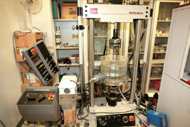 Staffed by one technician, this lab is equipped with a MTS Mini-Bionix bi-axial test system. The full range of mechanical testing can be performed for all regions of the human body (e.g. spine, hip and finger), following ASTM or ISO, all types of animals (e.g. goat, rabbit and mouse), as well as implants, protheses and biomaterials. Hip and spine kinematics simulations are also available with the specially-designed fixtures. In addition, the laboratory has the implant wear simulation testing system for biomechanical testing of small joints.
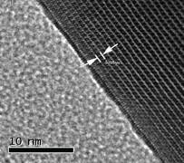
TEM photo of Sr-HA nanowhisker
The Biomaterials Laboratory
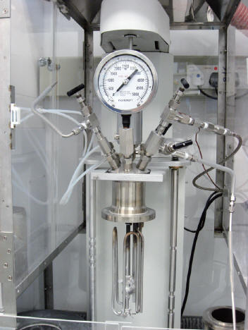
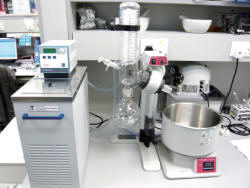 This lab develops biomaterials, bioactive bone cements, bio-microsphere for drug and cell delivery, and supply scaffolds for tissue regeneration and engineering. The lab is equipped with a ball mill grinder, sieve shaker, vacuum pump and apparatus, high temperature oven, etc. Of note is our recent success to develop a novel bioactive bone cement for prosthesis and implant fixation and treatment of osteoporotic fractures. The lab has also installed from small 100ml Teflon lined reactor to Parr 4547 large scale hydrothermal reactor for synthesis nano powder, nano rods in all kinds of biomaterials.
The Molecular and Cell biology Laboratory
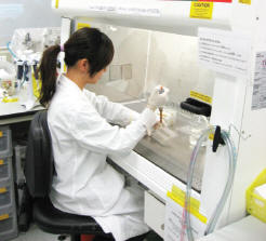
Molecular and cell biology laboratory
Most research is carried out in collaboration with the Department of Biochemistry, and many facilities and equipment are shared. Additionally the Genome Centre of the Li Ka Shing Faculty of Medicine provides core facilities and support for genomics and proteomics projects. Our relatively small laboratory received set-up funding from the HKU Foundation for development into an adult stem cell research laboratory, supporting work on musculoskeletal tissue regeneration. Projects such as treatment of osteoporotic fractures by encapsulated allogenic mesenchymal stem cells, intervertebral disc regeneration are underway.
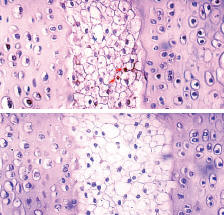
DNA-labeling retaining cells in the mouse intervertebral disc
The Histology and Experimental Surgery Laboratory
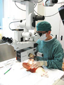
Histology and experimental surgery laboratory
This lab has all the facilities for performing standard histology on soft tissues and decalcified sections. Also an in-vivo micro CT scanner (Skyscan 1076) has newly arrived and installed for small animal in-vivo imaging. We have purchased a cutting and grinding system for hard tissues, to permit undecalcified histological sections to be performed. The other halve of the lab has been set up to perform animal surgery, up to the size of rabbits. In addition to standard operating room equipment, there is an operating microscope, a very useful tool for small animal surgery, as well as for practicing surgeons to sharpen their micro-dissection and repair skills.
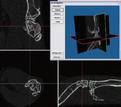
Three orthogonal reconstr ucted slices
of mouse knee, 8.8µm pixel size
of mouse knee, 8.8µm pixel size

Micro CT for live animals
Orthopaedic Skills Laboratory
Approximately 900 sq ft. is set aside for a skills laboratory and seminar room. It is intended that this lab will be used for cadaveric and saw-bone workshops, as well as for holding small to medium-sized seminars. We have custom-built seven removable workstations, each with compress air, vacuum suction, electricity and water supply. The laboratory aims to provide the highest standard technology to train orthopaedic surgeons locally and regionally in the following areas:
- Arthroscopic surgery
- Fracture fixation
- Hand Surgery
- Joint replacement surgery
- Limb lengthening Surgery
- Micro-surgery
- Spinal surgery
3D Bio-Printing
3D bio-printing is currently a hot topic in the field of research, particularly its applications in the medical field. To strengthen ourselves in this area, we recently bought a 3D Bioprinter, namely “3D Discovery” by regenHU, in O&T department. The “3D Discovery” is a versatile and cell friendly instrument. It can create three-dimensional models that more closely mimic what happens in living organisms. It allows scientists to pattern cells, biomolecules, and a range of soft and rigid materials in desirable 3D composite structures.
- The printer offers a printable area (W x L x H) of 130 x 90 x 60mm, with an axis motor resolution of 0.0025mm, and repeatability and accuracy per axis of less than 0.05mm.
- The “3D Discovery” offers a high modularity, allowing users to customize the printer for their specific objectives. The three distinct printing heads can be customized in order to offer their own specific applications.
- The “Time-Pressure” direct dispenser allows directing dosing out of the cartridge. This is typically used for viscous materials such as hydrogels. A UV curing component is also equipped on this printing head.
- The “Polymer Extruder” is used for the materials such as high-temperature polymers, which are not dispensable simply by pressure. This printing head incorporates a screw design to extrude the materials out and then form the scaffolds desired. The melting temperature can go up to 250oC.
- The “Cell Friendly” printing head is used for the materials with low to mild viscosity such as culture medium. It offers jetting as well as contact dispensing depending on the tip installed. A micro valve mechanism is used to control the dispensing of the media. The whole printer is located inside a class II biosafety cabinet, making cell-printing completely feasible. The cell-scaffolds printed can directly apply for animal implantation.
Duchess of Kent Children’s Hospital

The Clinical Electrophysiology and Neural Engineering Laboratory
Clinical electrophysiology & neural engineering
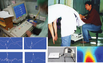
Intra-operative Spinal Monitoring – Two Nicolet Viking IV Electrophysiologic Systems and one Magstim-250 magnetic nerve stimulator were set-up in the operation theatres for spinal cord intra-operative monitoring and for research applications. Various evoked potentials detection and EMG Muscular Evaluation equipment are also located in this laboratory. Multi-channel functional electrical stimulator supported with orthosis was developed in this laboratory for paraplegic walking training. A dynamic three-dimensional forces and movements detection system has been developed to evaluate the performance of paraplegic walking under FES. The laboratory also has a SensorMedics 6200 Autobox DL whole body plethysmograph instrument for a complete, rapid and accurate measurement of lung function.
The Motion Analysis Laboratory
The motion analysis laboratory
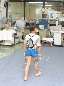
The laboratory is equipped with six cameras Vicon 370 3-dimensional motion analysis system, which is used in studying motions in the spine, peripheral joints and gait analysis. This laboratory is also equipped with an telemetric electromyography (EMG) machine, Kistler force platform and other computer equipment. As a result, both information on kinetic and muscle action can be studied.
Queen Mary Hospital

The Audio-Visual Laboratory
 Located in Queen Mary Hospital, this lab provides support for documentation of clinical cases in everyday service, and for publication of research work. It is equipped with digital photography, video and scanning support, video editing facilities, large size colour plotter for printing of posters, and darkroom facilities. Two laboratories are also located at the Duchess of Kent Children’s Hospital for the convenience of the patients.
The Tam Shiu Anatomical Modelling Laboratory
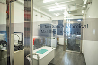 We provide clinical and research support consultation and production service in medical 3D printing. Established in 2018, the laboratory now provides service to more than 300 patients each year with complex pathologies requiring specialized 3D digital technology for intervention planning. The service is doctor led with an emphasis on rapid turnover, clinical relevance, quality, and clinical validation mandated in the production workflow. We encourage utilization of this facility for all pathoanatomically complex structural diseases from routine cases to the most unusual and innovative clinical situations. We offer specialist led service for orthopaedic cases which typically cover 60% of our load and collaborative consultation, design, and production for all other medical specialities in the remaining 40% of cases.
Our scope of service covers:
- Image segmentation and 3D modelling
- Patient specific 3D models for visualization of disease pathoanatomy
- Patient specific instruments
- Teaching and illustrative models
- Medical devices innovation
- Designing of patient specific implants
The anatomical modelling lab is supported by a generous donation from the Tam Shiu Charitable Foundation and HKU research funding. Our mission is to provide rapid quality service while minimising financial burden to entitled users. Our primary service target includes:
- All Hong Kong public and private hospitals
- Orthopaedic and all medically related specialities
- Allied health
- Medical devices innovation partners
- Collaboration with other academic units
Our primary production capability includes:
-
- Polymer Fused deposition modelling– single material ABS-M30i or Ultem 1010 (Stratasys)
a. For low cost, rapid turnover services
b. Simple anatomical models
c. Surgical Jigs and fixtures
d. Functional devices
-
- Polymer Selective laser sintering printer – single material PA-2200 (EOS)
a. High resolution and high durability models
b. Autoclavable surgical jigs and fixures
c. Functional devices
-
- Polymer Polyjetting – multiple materials (Stratasys J750 Digital anatromy)
a. Complex anatomical models
b. Parts requiring transparency, rubber like consistency, and multiple colours
c. Functional parts for training and medical device innovation
d. Rapid turnover
- Metal – Outsourcing to industrial partner
For request of service and collaboration, please contact our secretary for more information: manfai@hku.hk
MacLehose Medical Rehabilitation Centre

Young Chi Wan Bone & Joint Research laboratory
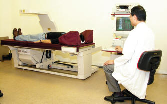
Bone densitometry
This laboratory is in the MacLehose Medical Rehabilitation Centre, where a dual-energy X-ray absorptiometry machine is installed to measure bone mineral density for both service and research. In particular, it is used to study bone remodeling around total hip prostheses.
Biomechanics laboratory
TEM photo of Sr-HA nanowhisker
Biomaterials Laboratory
Molecular and cell biology laboratory
DNA-labeling retaining cells in the mouse intervertebral disc
Micro CT for live animals
Three orthogonal reconstr ucted slices of mouse knee, 8.8µm pixel size
Histology and experimental surgery laboratory
Bone densitometry
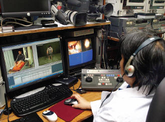
The audio-visual laboratory
Clinical electrophysiology & neural engineering
The motion analysis laboratory