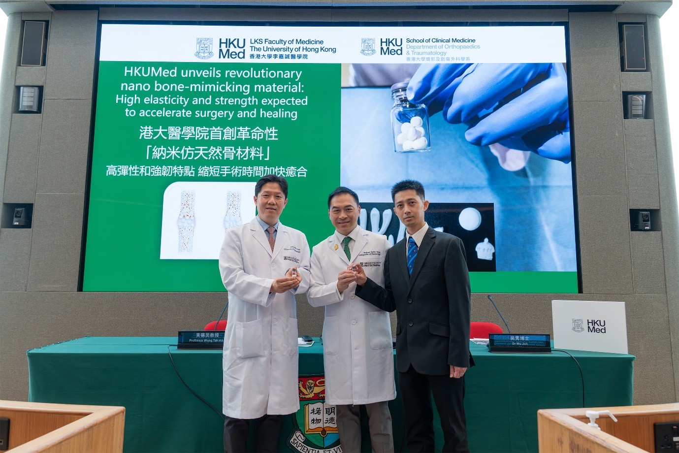
HKUMed develops an innovative titanium implant surface for rapid bacteria elimination and enhanced bone regeneration

A research team from the Department of Orthopaedics and Traumatology, School of Clinical Medicine, LKS Faculty of Medicine at the University of Hong Kong (HKUMed), has developed a titanium implant surface that can be activated by near-infrared (NIR). With just 15 minutes of NIR irradiation, this surface can eliminate 99.94% of Staphylococcus aureus (S. aureus) biofilms without the use of antibiotics, while simultaneously promoting bone-implant fusion. Based on titanium dioxide (TiO2), the same compound found in titanium’s natural surface oxide layer, the design may offer practical advantages for compatibility with existing titanium implants and future clinical translation. This innovative technology could be applied to various common orthopaedic implants, including joint replacements, fracture fixation devices, spinal fusion cages, dental implants and craniofacial implants. It offers a new solution to combat implant infections. The findings were published as a cover story in the international journal Cell Biomaterials [link to the publication].
Implant-associated infections remain a major clinical challenge in orthopaedic practice. Once bacteria adhere to an implant surface and form biofilms, they become highly tolerant to antibiotics and can evade the patient’s immune system. Consequently, patients often require repeated invasive debridement, revision surgeries and prolonged courses of high-dose systemic antibiotics. These treatments extend recovery time, increase healthcare costs, contribute to antibiotic resistance and often fail to prevent recurrent infections.
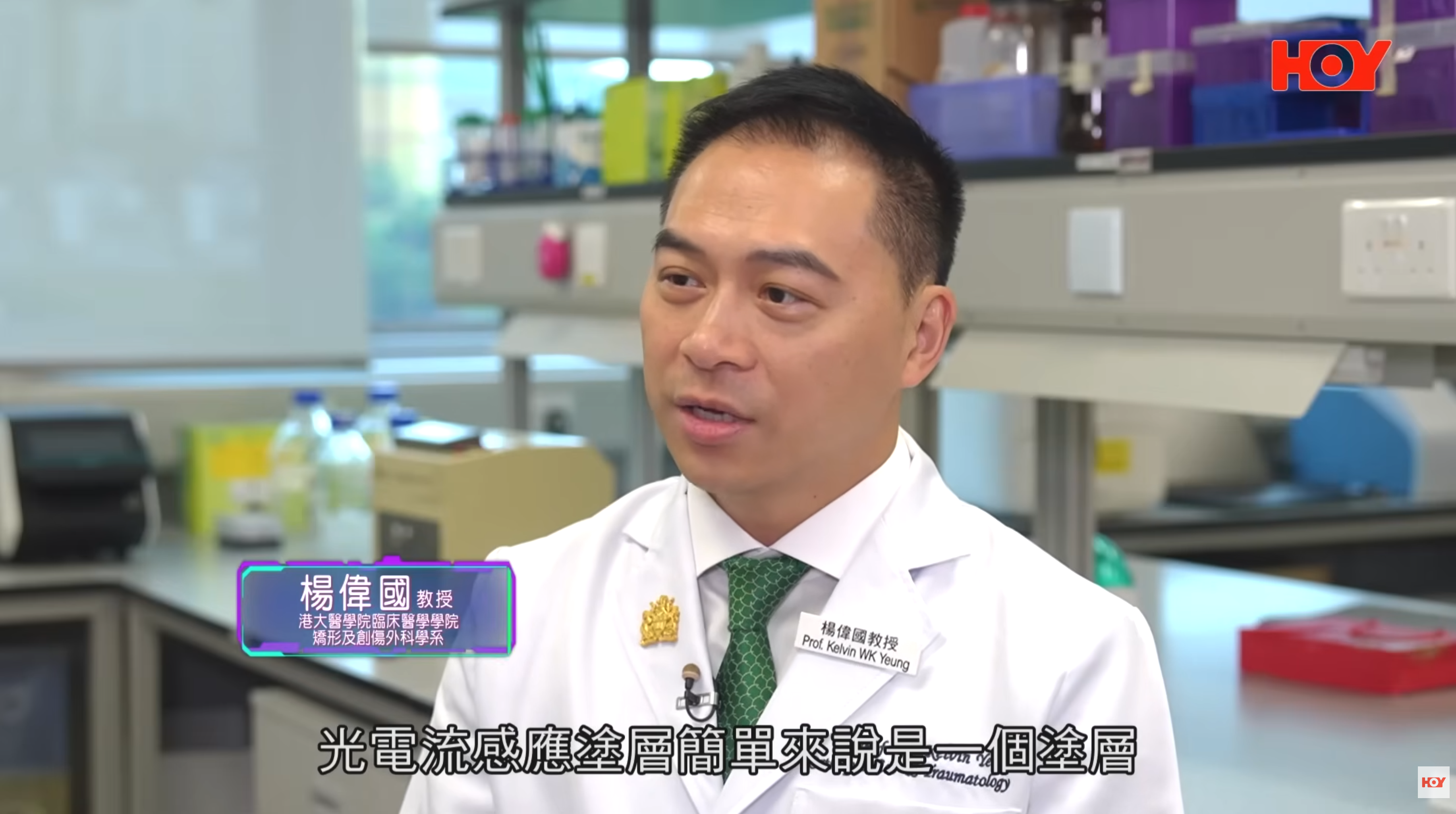
Professor Kelvin Yeung Wai-kwok, from the Department of Orthopaedics and Traumatology, School of Clinical Medicine, HKUMed, who led the research, stated, ‘Implant-associated infection is a major cause of implants failure. Bacterial colonisation and biofilm formation on implant surfaces can be difficult to eradicate, often resulting in persistent inflammation, compromised implant fixation and, ultimately, loosening and failure.’
‘Although existing antibacterial coatings can provide some level of protection, they typically rely on loaded agents such as antibiotics, metal ions or complex bioactive molecules,’ he added. ‘Their effectiveness, however, is limited by their loading capacity, uncontrolled release, potential cytotoxicity and loss of function once these agents are exhausted. These limitations underscore the urgent need for an in situ, controllable and durable strategy that can both eliminate biofilms and promote bone-to-implant integration without the use of additional drugs or agents. This is precisely what our research seeks to achieve.’
Light-triggered defence enables rapid bacterial elimination
Currently, titanium and titanium alloys, which are widely used in orthopaedic surgery, naturally form a very thin TiO2 layer on the implant surface. While this layer is highly biocompatible, it offers minimal defence against infection and lacks the ability to actively promote bone growth. When bacteria such as S. aureusform biofilms on these surfaces, antibiotics are often unable to eradicate the infection effectively, resulting in the need for invasive revision surgeries.
Using a customised template-assisted technique, the HKUMed team precisely engineered nano-honeycomb structures on the titanium surfaces and introduced oxygen vacancies through hydrogenation treatment, creating a remotely activatable smart surface. Under NIR irradiation, this smart surface generates reactive oxygen species and a mild local photothermal effect, rapidly disrupting biofilm architecture and killing bacteria.
In vitro experiments found that a single 15-minute irradiation was sufficient to eliminate 99.94% of S. aureus. In a rat tibial defect infection model, the same treatment removed 91.58% of biofilms. Compared to unmodified titanium implants, the engineered surface can efficiently perform biofilm clearance and lead to marked reductions in pus formation and local inflammation, resulting in a notable increase in new bone formation around the implant.
Enhancing the osteoimmune microenvironment supports new bone formation
Beyond its strong antibacterial performance, the research demonstrated that the new implant surface effectively modulates the local immune response. It shifts macrophages from a prolonged pro-inflammatory state to a pro-healing, tissue remodelling phenotype, thereby creating a more favourable osteoimmune microenvironment. This attracts more osteogenic cells and facilitates their differentiation, leading to a significant increase new bone formation. As a result, the implant achieves faster and more stable integration with the bone, demonstrating that the new technology prevents infection while accelerating bone fusion.
‘By adopting an agent-free strategy, our newly developed smart surface demonstrates remarkable antibacterial and pro-osteogenic capabilities without the use of additional drugs,’ explained Professor Yeung. ‘This technology offers high translational potential, as it relies solely on native implant materials and mature manufacturing processes. This will facilitate regulatory approval, support scalable manufacturing and pave the way for future clinical adoption. We believe this innovation will broadly improve orthopaedic surgical outcomes and benefit more patients.’
About the research team
The research study was led by Professor Kelvin Yeung Wai-Kwok, Department of Orthopaedics and Traumatology, School of Clinical Medicine, HKUMed. The first author is Dr Zhu Yizhou, Research Assistant Professor in the same department.
Acknowledgments
The research is jointly supported by funding from the Chinese Mainland, including the National Key Research and Development Programme of China, the National Natural Science Foundation of China, the Shenzhen Science and Technology Innovation Commission, the Guangdong Basic and Applied Basic Research Foundation, and the National Science Fund for Distinguished Young Scholars; as well as the General Research Fund, the Collaborative Research Fund, the Hong Kong Innovation Technology Fund, the Health and Medical Research Fund, the National Natural Science Foundation of China/ RGC Joint Research Scheme, the Government of the Hong Kong Special Administrative Region of the People’s Republic of China.
港大醫學院首創智能鈦金屬植入物塗層 迅速殺菌兼促進新骨生成融合
2026年4月1日
香港大學李嘉誠醫學院(港大醫學院)臨床醫學學院矯形及創傷外科學系的研究團隊,成功研發出一種可由近紅外線啟動的鈦金屬植入物塗層,只需以近紅外線單次照射 15 分鐘,即可快速清除金黃葡萄球菌生物膜達到 99.94%,更無需使用抗生素,亦能同時促進骨骼與植入物的融合。該設計以二氧化鈦為基礎,而二氧化鈦亦是鈦金屬天然表面氧化層中的相同化合物,這一特點或許為其與現有鈦金屬植入物的配合應用及未來臨床轉化帶來優勢。這項技術有望應用於多種常見骨科植入物,包括關節置換、骨折內固定裝置、脊椎融合器、牙科植體及顱顏植入物等,為植入物感染問題提供全新的解決方案。相關研究成果已刊於國際期刊《Cell Biomaterials》,並成為封面故事(按此瀏覽期刊文章)。
植入物相關感染是骨科手術的一大挑戰。當細菌於植入物表面形成生物膜,便會對抗生素及患者的免疫系統產生耐藥性。患者往往需要接受反覆清創、翻修手術和長期使用高劑量全身性抗生素,這些治療程序延長了康復時間、增加醫療開支,亦可能加劇抗生素耐藥風險,以致出現反覆感染。
領導此研究的港大醫學院臨床醫學學院矯形及創傷外科學系教授楊偉國教授表示:「植入物相關感染是導致植入物失效的重要原因。細菌一旦在植入物表面產生並形成生物膜,通常難以清除,並往往會引致持續發炎,令植入物未能固定,最終造成鬆脫甚至失效。」
楊偉國教授續指:「現有抗菌塗層雖能提供一定保護,但多依賴外加藥劑,如抗生素、金屬離子或複雜生物活性分子。這些方法載藥量有限,釋放劑量也難以精準控制,還可能有潛在毒性,而且藥效消退後抗菌功能便會隨之消失。因此,臨床上亟需一種新技術,可在體內原位啟動、效果可控持久,既能高效清除生物膜,又能促進骨骼與植入物的融合,而無需使用額外藥物。這正是我們的研究方向。」
光熱效應迅速殺菌
現時廣泛應用於骨科手術的鈦金屬及其合金植入物塗層,具有一層天然形成的二氧化鈦薄膜,它具有良好生物相容性,但缺乏主動抗菌和促成骨骼生長的能力。當金黃葡萄球菌等細菌在鈦塗層上形成生物膜後,單靠抗生素難以根除細菌,令患者常需接受侵入性翻修手術。
港大醫學院團隊以模板輔助技術,在鈦塗層精準構建納米蜂窩狀結構,進行氫化處理引入氧空位,製成可遙距啟動的智能塗層。在近紅外線照射下,這種智能塗層可同步產生活性氧和溫和的局部光熱效應,迅速破壞生物膜結構並殺滅細菌。
在體外實驗中,單次 15 分鐘照射智能塗層,即可清除 99.94% 金黃葡萄球菌;在大鼠脛骨缺損感染模型中同樣能消除 91.58% 的生物膜。與傳統鈦植入物相比,智能塗層可顯著減少生物膜殘留、膿液形成及局部炎症反應。同時,該塗層有效調節免疫反應並促進骨骼生長,顯著提升植入物周圍的新骨形成。
優化骨骼免疫微環境助新骨生成
除了強效的抗菌性能外,研究亦證實這種新型植入物塗層能有效調控免疫反應。它可促使巨噬細胞由持續的「促炎狀態」,轉向有助組織修復的「促癒合狀態」,改善局部組織修復環境,吸引更多成骨細胞並促進其分化,明顯有助新骨生成,令植入物更快、更穩定地與骨骼融合。這證明了新技術不但能抗感染,亦可加速骨融合,彌補現有植入物的不足。
楊偉國教授表示:「我們研發的新型植入物塗層在『不添加藥劑』的情況下,不僅展現出強效的抗菌和促成生骨能力,同時具備極高的轉化潛力。這項技術完全依賴現有植入物材料,製造流程成熟,有利於監管審批、規模化生產及臨床應用,未來將可廣泛改善骨科手術成效,惠及更多患者。」
關於研究團隊
這項研究由港大醫學院臨床醫學學院矯形及創傷外科學系教授楊偉國教授領導。第一作者為同學系的助理教授(研究)祝亦周博士。
鳴謝
此項研究獲得多項資助支持,包括國家重點研發計劃、中國國家自然科學基金、深圳市科技創新委員會、廣東省基礎與應用基礎研究基金,以及國家傑出青年科學基金;中華人民共和國香港特別行政區政府研究資助局優配研究金及協作研究金、創新科技署創新及科技基金、醫務衞生局醫療衞生研究基金、國家自然科學基金委員會及研究資助局聯合科研資助基金等。